Молекулярные модели ДНК и тРНК

На схеме представлены вандерваальсовые модели (см. Строение молекул) двух небольших нуклеиновых кислот. На схеме А изображена ДНК (DNA), построенная из 17 пар нуклеотидов, на схеме Б — структура фенилапанинспецифичной тРНК (tRNA) дрожжей (75 нуклеотидов).
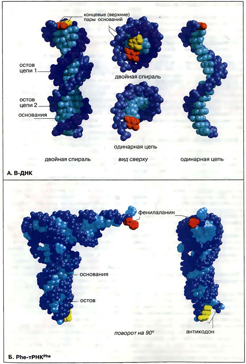

Биомолекулы. Нуклеиновые кислоты / Молекулярные модели ДНК и тРНК

Статьи раздела «Молекулярные модели ДНК и тРНК»:

Следущая статья   |   — Вернуться в раздел


Микробиология / В учебнике изложены основы современной микробиологии, вирусологии и иммунологии. Учебник состоит из двух частей. Первая часть — общая микробиология — включает основные сведения о морфологии, физиологии микроорганизмов, антибиотиках, генетике бактерий, инфекции, иммунитете и аллергии. Вторая часть — Микробиология
В учебнике изложены основы современной микробиологии, вирусологии и иммунологии. ...
Gene Control / Gene Control offers an articulate, current description of how gene expression is controlled in eukaryotes, reviewing and summarizing the extensive primary literature into an easily accessible format. Gene Control will be of value to students in biological sciences, as well as to scientists and cliniGene Control
Gene Control offers an articulate, current description of how gene expression is controlled in eukaryotes, reviewing and summarizing the extensive ...
Stress — From Molecules to Behavior: A Comprehensive Analysis of the Neurobiology of Stress Responses / This book comprehensively covers the molecular basis of stress responses of the nervous system, providing a unique and fundamental insight into the molecular, physiological and behavioral basis of the stress response of a whole organism. Edited by leading experts in the field and summarizing the latStress — From Molecules to Behavior: A Comprehensive Analysis of the Neurobiology of Stress Responses
This book comprehensively covers the molecular basis of stress responses of the nervous system, providing a unique and fundamental insight into the ...
NMR Studies of Structural Motifs: Protein Folding and Ligand Binding / NMR of Structural Motifs: The agrin G3 domain is critical in development and maintenance of the neuromuscular junction. G3 binds -dystroglycan and initiates acetylcholine receptor clustering on myotube membranes. Using NMR spectroscopy, we show both active B8 and inactive B0 isoforms binding sialic NMR Studies of Structural Motifs: Protein Folding and Ligand Binding
NMR of Structural Motifs: The agrin G3 domain is critical in development and maintenance of the neuromuscular junction. G3 binds -dystroglycan and ...